Helen King on Structure Evaluation Susan Garrett's Dog Training Blog
Summary Anatomy of a Dog Dog anatomy details the various structures of canines (e.g. muscle, organ and skeletal anatomy). The detailing of these structures changes based on dog breed due to the huge variation of size in dog breeds. Would you be surprised to know that short dogs are more aggressive? Or taller dogs are more affectionate?

FileDog anatomy lateral skeleton view.jpg
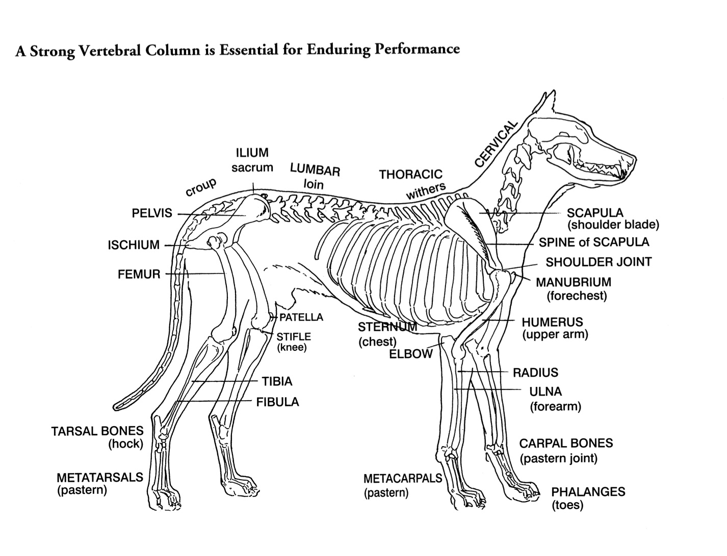
This is an overview of the canine skeleton, pointing out features that help in identification and orientation.If you find this helpful, please let me know by.

Anatomy Of Dog Skeleton With Labeled Inner Bone Scheme Vector
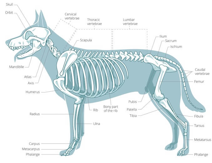
Here are presented scientific illustrations of the canine skeleton, with the main dog's bones and its structures displayed from different anatomical standard views (cranial, caudal, lateral, medial, dorsal, palmar..). Some of the different canine joints are labeled.

Vintage 1935 Dog Veterinary Print Skeleton Of Dog Anatomy Of Dog Canine
The dog skeleton is the bony part of dogs made for the support and protection of internal organs. Bones are connected through joints and muscles move the bones to produce the normal dog movements. In this article we will cover: Bone types and parts of the dog skeleton The dog skull Dog cranium The spine The Trunk The Forelimb The Hindlimb
A Visual Guide to Dog Anatomy (Muscle, Organ & Skeletal Drawings) All
An overview of the anatomy of the canine skeleton.Follow on twitter @ https://twitter.com/PerkyVetInstagram: Perkydvm

Dog skeleton 101 Dog Anatomy Bones Animal Hackers
Components of the Musculoskeletal System in Dogs. Bones provide rigid structure to the body and shield internal organs from damage. They also house bone marrow, where blood cells are formed, and they maintain the body's reservoirs of calcium and phosphorus. Old bone tissue is constantly replaced with new bone tissue in a process called.
Canine Skeleton Poster Clinical Charts and Supplies
Labeled anatomy of the head and skull of the dog on CT imaging (bones of cranium, brain, face, paranasal sinus, muscles of head) This module of vet-Anatomy presents an atlas of the anatomy of the head of the dog on a CT. Images are available in 3 different planes (transverse, sagittal and dorsal), with two kind of contrast (bone and soft tissues).
Dog Skeletal Skull Anatomy Poster 18 X 24 Etsy
This veterinary anatomical atlas includes selected labeling structures to help student to understand and discover animal anatomy (skeleton, bones, muscles, joints, viscera, respiratory system, cardiovascular system). Positional and directional terms, general terminology and anatomical orientation are also illustrated.

Dog Anatomy Dog Skelton
A dog's physical anatomy is designed to help them navigate their environment and perform various tasks. Their bodies are made up of many different parts, including their skeleton, muscles and internal organs. One of the most important parts of a dog's anatomy is their skeleton.

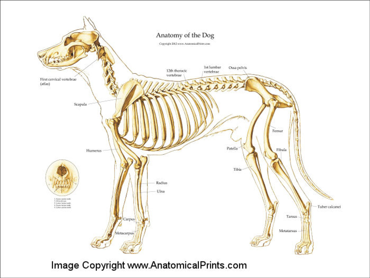
Dog skeleton with major bone elements labeled (Davis, 1987, p. 54
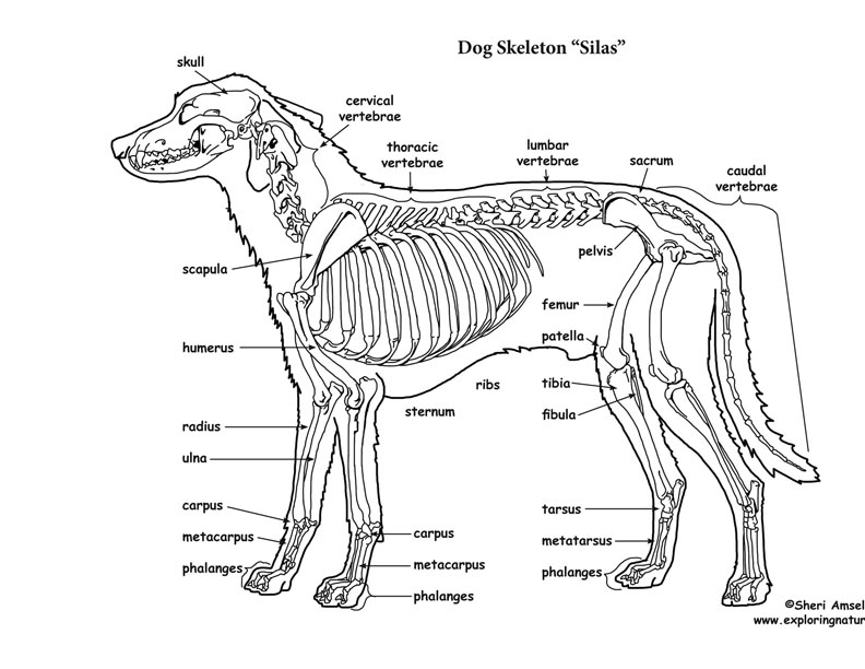
January 3, 2024 < http://www.exploringnature.org/db/view/Dog-Skeletal-Anatomy > Dog Skeletal Anatomy
Anatomy Of Dog Skeleton With Labeled Inner Bone Scheme Vector
Dog anatomy comprises the anatomical studies of the visible parts of the body of a domestic dog.Details of structures vary tremendously from breed to breed, more than in any other animal species, wild or domesticated, as dogs are highly variable in height and weight. The smallest known adult dog was a Yorkshire Terrier that stood only 6.3 cm (2.5 in) at the shoulder, 9.5 cm (3.7 in) in length.
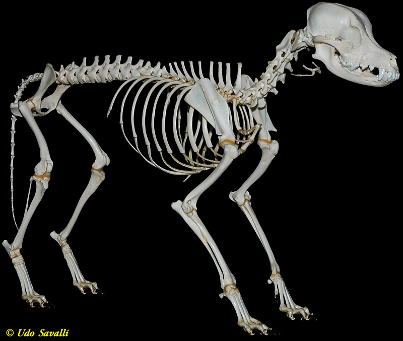
BIO370Mammal Skeletons
Organs of dogs Canine anatomy As we explain above, canine anatomy is far ranging due to the diversity of existing breeds. These different breeds not only differ from each other in size, but in the shape of many body parts. Perhaps the most significant is head shape. There are three main different types of head formation in dogs:
Dog Skeleton Anatomy
The cat has a small coronoid fossa medial to the radial fossa that accommodates the coronoid process of the ulna during elbow joint flexion.; The cat has a supracondylar foramen near the medial condyle allowing the passage of the median nerve and brachial blood vessels.; There is an intermediate tubercle between the greater and lesser tubercles in the horse's intertubercular groove.

maryrosedog Dog skeleton, Dog anatomy, Dog skull
The skeleton is composed of the hard tissues of the body, and its primary functions are to support the body, to provide a system of levers used in locomotion, to protect the soft organs of the body, and to produce red blood cells (hematopoiesis). A dog's skeleton is formed so the dog can run fast, hunt and chase.
Dog Skeletal Anatomy
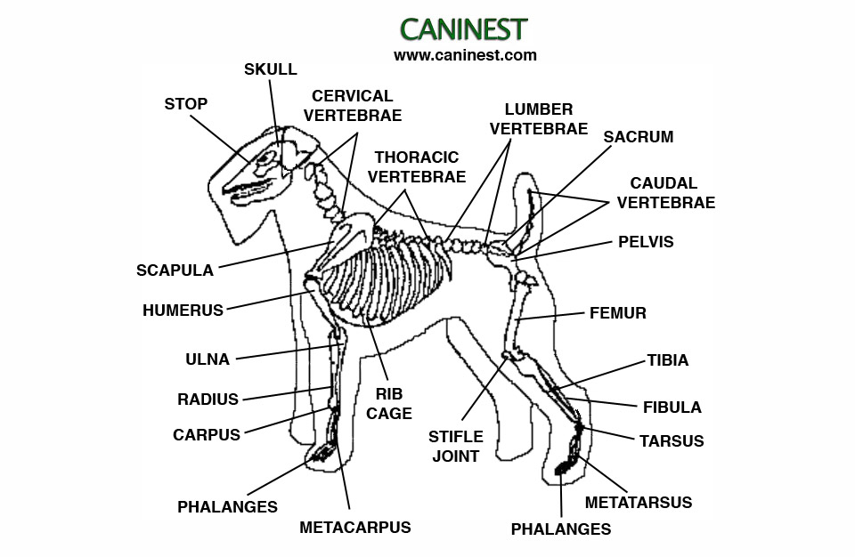
Directional Terms and Anatomic Planes Directional Terms from Normal Stance (Anatomic Position) The dog stands upright on digits or phalanges of each forepaw or manus and each hindpaw or pes (Figure 5-1). This type of stance is termed a digitigrade stance.

Dog skeleton 101 Dog Anatomy Bones Animal Hackers
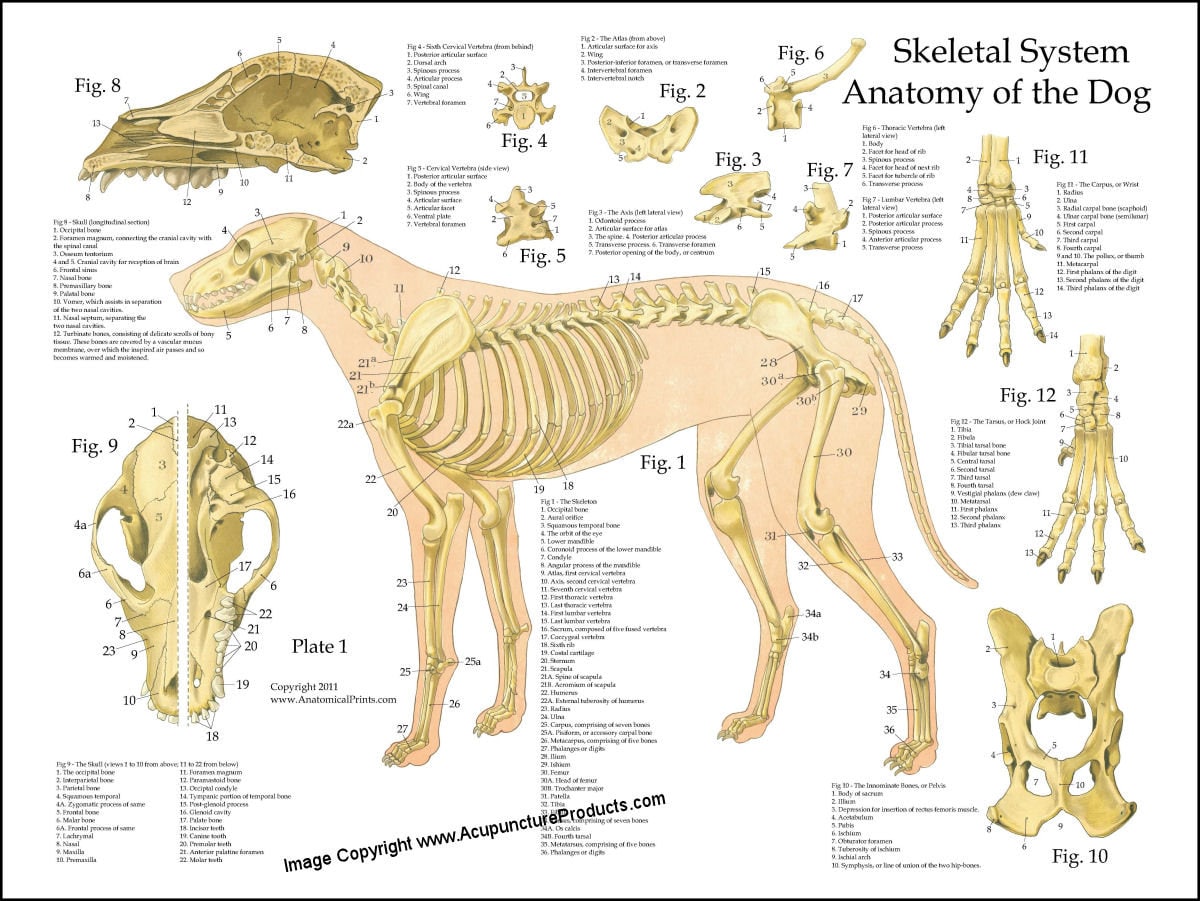
Dog skeleton, TBjornstad, 2006 The information below will highlight the bone anatomy of the domestic dog which will include the major and selected important minor aspects of anatomy. Very detailed anatomy of individual bones is not covered below due to space restrictions.